Nonunion Fractures Treatment
A nonunion fracture is a disruption in the fracture repair process. Bones possess a remarkable ability to heal themselves, and with appropriate treatment, most fractures will heal without complications. However, due to certain factors, such as inadequate stability or poor blood supply, some fractured bones fail to heal properly and do not reunite within the expected timeframe, typically six to nine months post-injury. Unlike standard fractures that progress through the normal healing stages, nonunion fractures remain unhealed, leading to prolonged pain and functional impairment.
Treatment in Cuba for nonunion fracture is dependent of the underlying cause. Only once the cause of the nonunion is identified can proper treatment be initiated. However, If the nonunion is out of alignment, surgery will be necessary to realign and stabilize the bones. Should the nonunion fracture not be treated, it can lead to:
- Chronic pain
- Functional impairment
- Deformity
- Increased risk of further injury
- Potential severe complications such as infection or osteoarthritis in the affected area
Types of Nonunion Fractures
There are several types of nonunion fractures classified according to the characteristics of the fracture site.
- Atrophic Nonunion: Caused by inadequate immobilization and inadequate blood supply, resulting in no callus formation.
- Hypertrophic Nonunion: Caused by inadequate stability with adequate blood supply. There is callus formation, but the bone ends are not aligned properly, preventing healing.
- Oligotrophic Nonunion: Often results from inadequate stabilization of the fracture with fracture fragment displacement with insufficient callus formation.
- Infected (Septic) Nonunion: Results from an infection at the fracture site, leading to poor healing despite biological activity.
- Pseudoarthrosis: A false joint, or pseudoarthrosis, forms at the fracture site due to excessive movement and inadequate healing.
Causes of Nonunion Fractures
The primary factors contributing to nonunion include:
- Inadequate stabilization.
- Poor blood supply.
- Infection at the fracture site.
- Systemic factors (conditions such as osteoporosis, diabetes, smoking, and certain medications).
- Severe trauma or high-energy fractures.
- Nutritional deficiencies (such as calcium and vitamin D).
Symptoms of Nonunion Fractures
Patients with nonunions usually feel pain at the site of the break long after the initial pain of the fracture disappears. Other symptoms include:
- Swelling and tenderness that do not subside over time.
- Visible deformity or instability in the affected area.
- Limited range of motion or functional impairment.
- Absence of radiographic evidence of bone healing over time.
Diagnosing of Nonunion Fractures
Diagnosing nonunion fractures involves a comprehensive evaluation combining clinical assessment and imaging techniques to determine the exact nature and extent of the nonunion.
- Medical History: This includes history of the original injury, previous treatments, and any underlying conditions (such as diabetes, smoking, or osteoporosis) that could affect healing.
- Symptoms Review: Discussion of symptoms such as persistent pain, swelling, tenderness, and functional impairment that have persisted beyond the normal healing period.
- Physical Examination: Visual examination of the affected area, palpation and functional tests.
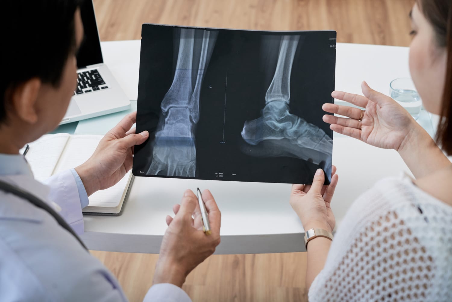
- X-Rays: Usually, the first imaging modality used to assess bone alignment, the presence of callus formation, and overall bone healing.
- Computed Tomography (CT) Scan: These are detailed cross-sectional images of the bone and surrounding tissues used to identify subtle fractures, bone gaps, and nonunion characteristics. Additionally, they are used to evaluate the extent of bone bridging and any potential hardware complications from previous surgical interventions.
- Magnetic Resonance Imaging (MRI): These images are used to assess the condition of the soft tissue surrounding the fracture and to identify signs of infections or inflammation.
- Ultrasound: These images are used to assess callus formation
- Blood Tests: Blood tests are used to help identify infection or inflammation and to assess nutritional status.
Treatment Options for Nonunion Fractures
Treating nonunion fractures is complex and multifaceted, often requiring a combination of surgical and nonsurgical approaches. Treatment option depends on several factors, including:
- Type of nonunion.
- Location of the fracture.
- Duration of nonunion.
- Previous treatments and to what result.
- Presence of comorbidities.
- Patient’s age.
- Patient’s overall health.
- Patient’s activity level.
Non-Surgical Options
- Electrical bone stimulation: This involves applying electrical currents to the fracture site with the aim of promoting bone growth and accelerating the healing process.
- Fracture brace immobilization: This type of brace is used for support to stabilize the bone, allowing for controlled movement and gradual weight-bearing to stimulate healing.
Surgical Options
- Fracture Fixation: Fracture fixation is a surgical procedure performed to stabilize a broken bone, and to ensure proper alignment and support as it heals. This procedure often involves the use of metal hardware, such as screws, rods, or plates, to hold the fractured bone segments in place. Fracture fixation could be internal or external.
- Internal fixation stabilizes a nonunion with metal plates and screws to the outside of the bone or places a nail (rod) inside the bone to hold the bone in position.
- External fixation stabilizes the injured bone by attaching a rigid frame to the outside of the injured arm or leg. The frame is attached to the bone with wires or pins. External fixation can treat nonunions in a patient who also has bone loss and/or infection.
- Debridement: Debridement is the process of removing dead, damaged, or infected tissue from a wound to promote healing. This procedure is essential for preventing infection, reducing inflammation, and creating a healthy environment for new tissue to grow.
- Soft tissue coverage: This procedure is utilized particularly when the surrounding tissue is compromised. Various surgical techniques, such as the use of soft tissue flaps, are used to cover and protect the fracture site to provide necessary blood supply and support to the healing bone
- Management of underlying conditions: This involves addressing systemic issues like infection, nutritional deficiencies (such as calcium and vitamin D), and chronic diseases (such as diabetes or thyroid disorders). Additionally, lifestyle modifications such as smoking cessation and limiting alcohol intake may be recommended.
PRIVATE ROOM WITH THE FOLLOWING FEATURES:
- Electronic patient bed
- Equipment for disabled patient
- Oxygen hookup
- Three AP meals taking into account the patient’s preferences and / or special diets prescribed by physician
- Fully equipped private bathroom
- Infirmary and nursing care
- Colour TV with national and international channels
- Local and international phone services (extra cost will apply)
- Safe box
- Internet service on every floor
- Laundry services
ADDITIONAL SERVICES INCLUDED IN THE PROGRAM:
- Assistance in visa issuance and extension (If needs be)
- Each patient/ companion will be assigned a multi-lingual field member with the mandate of attending to all of our patients’ translation and personal needs;
- 20 hours internet service;
- Local airport pickup and drop off; and
- Hospital pickup and drop off (if needed)
References :
–> WHY CUBA AS A MEDICAL TREATMENT DESTINATION
–> WHY CHOOSE CUBAHEAL